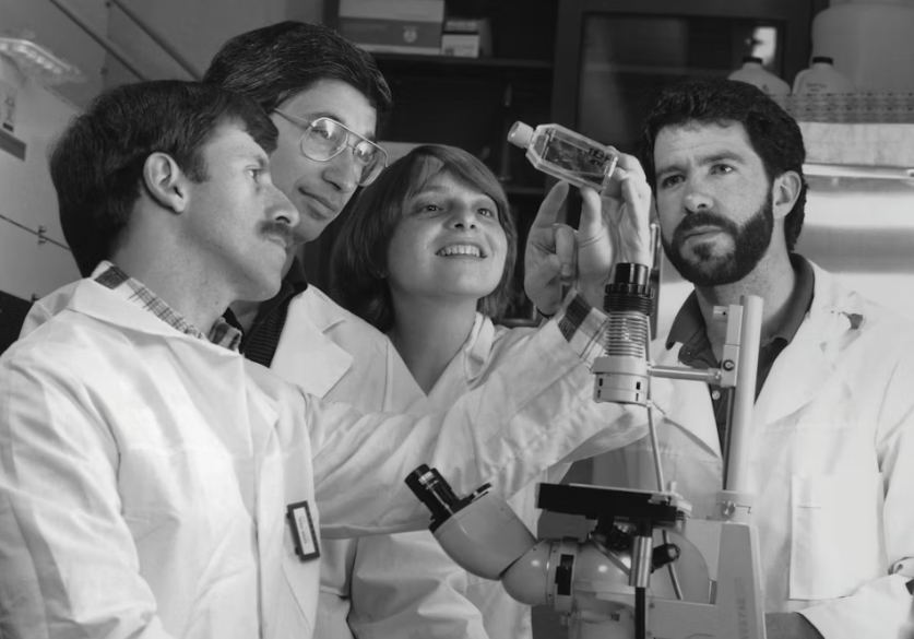
Few sciences can capture people’s imagination like palaeontology. Dinosaurs, mammoths, and other ancient organisms have a unique ability to leave an impression and excite ordinary people.
But palaeontology is an unusual, and rather odd, science. Palaeontologists are entirely dependent on new fossil finds to advance research, often leaving new discoveries down to fate more than most other areas of science. In palaeontology, fossils are more precious than gold.
How to extract a fossil
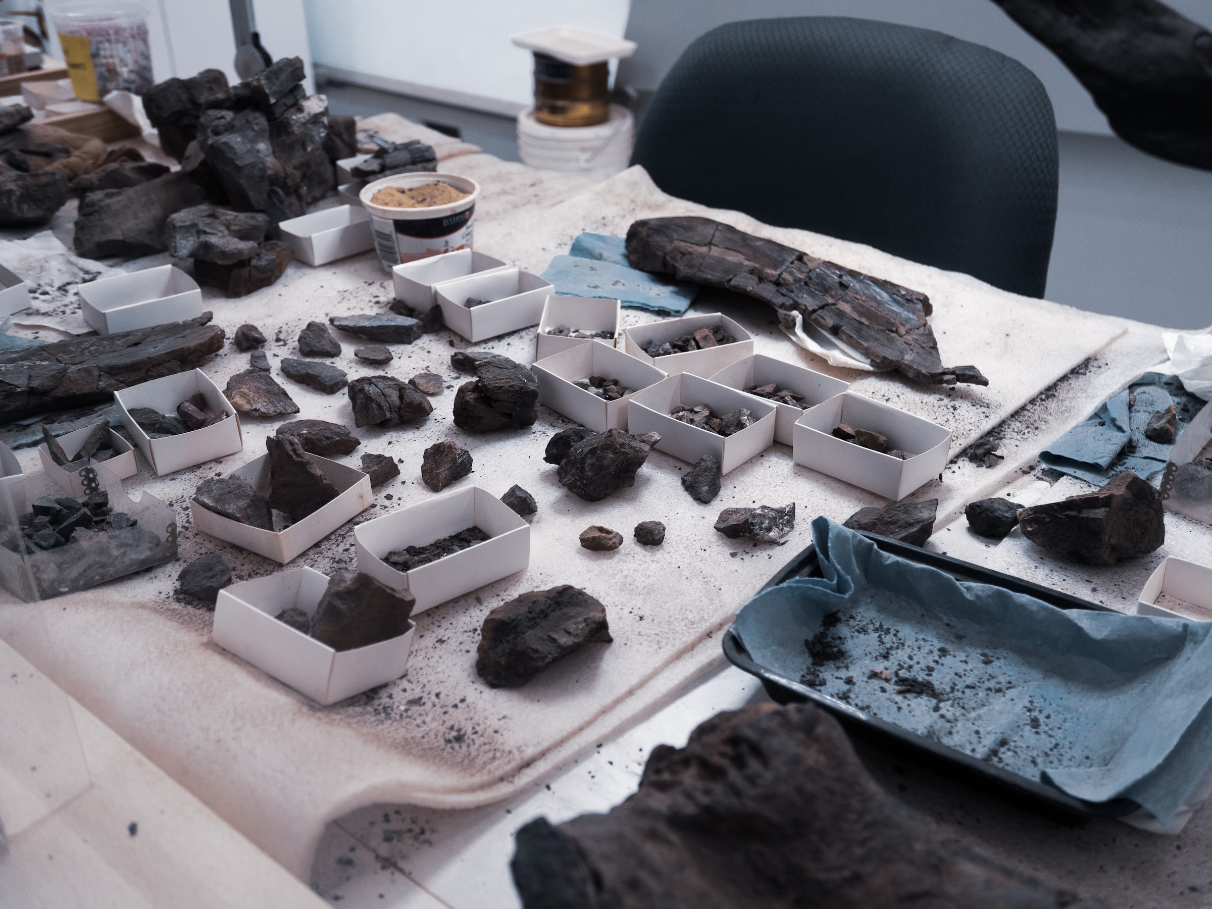
When a new fossil is found it is usually almost entirely encased in rock, known as the “matrix.” When the matrix covers so much of the fossil, a significant amount of useful information about the preserved organism is hidden.
Finding an effective method for revealing this elusive information about the fossils has been the aim of palaeontologists for centuries.
Complicating matters is an inherent trade-off between accessing the fossil and preserving its integrity during extraction, and the choice of methods with which to remove the matrix. In essence, the more matrix you remove the more of the fossil you can see, but the risk of damaging the fossil below the surface also then increases.
For larger fossils this is not such an issue—a few scratches may be barely noticeable. This has enabled the preparation, study, and display of the phenomenal dinosaur skeletons we are all familiar with, such as Dippy the Diplodocus at London’s Natural History Museum or Sue the Tyrannosaurus rex at the Chicago Field Museum (the largest T. rex ever found).
But for fossils of smaller animals, where a slip of the hand can break a bone or destroy a skull, removing the matrix is simply too risky.
I study the early evolution of life on land, looking at the very first creatures that grew legs instead of fins and dared to venture out of the water. Unlike the dinosaurs, most of these early tetrapods were incredibly small, sometimes just a few centimetres long.
As a consequence, the features used for identification and study are just millimetres long, or less. Gaining access to these fragile pieces of the Earth’s past is therefore like walking on a tightrope.
How to extract very small fossils
For 150 years, it seemed that this would be an insurmountable problem. Remove the rock, destroy the fossil. Leave it there and fail to uncover what is hidden beneath the surface.
In the early 1980s, Jim Elliot invented the first micro-CT scanner. CT scans use X-rays from many different angles to take thousands of X-ray photos, penetrating deep into objects, and allowing a 3D model to be constructed. They are best known for their use in medicine, where a patient lies still, and an X-ray-producing tube spins around them.
Elliot’s revolutionary technique was to have the X-ray-producing tube remain still, and the subject of the scan to rotate instead. This approach allows the X-ray-producing tube and camera to be much closer to the specimen, enabling a much higher dose of X-rays to be used. This achieves far greater image resolution.
The kind of X-ray dosage micro-CT scanners produce means they cannot be used for medical purposes—it is too dangerous for living subjects.
By the mid-2000s, micro-CT scanners had improved to the point where their resolution was just a few microns (where 1 micron equals 0.001 millimetres), and they were able to penetrate deep inside dense objects such as rock. This was the start of a revolution in the field.
Subsequently, studies emerged which utilised micro-CT scans to analyse fossils preserved in amber. Researchers produced remarkable 3D models of long dead insects, revealing them in greater detail than ever before.
Insects just a millimetre long, preserved from over 100 million years ago could have every bump on their bodies revealed in astonishing detail.
New ways of studying the old
Micro-CT scanners continued to improve in power and resolution, and by the start of the 2010s, micro-CT had become well-established as an effective method for peering deep inside rocks without compromising the integrity of the fossils within.
The 2010s saw a breath-taking flurry of new insights into fossilised organisms obtained through micro-CT scanning. It is hard to overstate just how much we have been able to learn as a result of this revolutionary technique.
Take, for example, Euryodus. Euryodus was an animal known as a “microsaur,” a small lizard-like animal that lived almost 300 million years ago and was one of the first vertebrates to live on land.
Euryodus fossils were first described in 1939, but historically scientists had always been unable to reveal details of its anatomy which are crucial in understanding how this animal lived and which others it was related to.
In 2020, Dr Bryan Gee and colleagues from the University of Toronto described Euryodus using micro-CT scans. They were able to produce a detailed 3D model of the skull, revealing every detail of the roof of the mouth, braincase, and jaws.
This revealed, for example, multiple rows of teeth in the lower jaws that may have been used for crushing the tough exoskeletons of insects. This morphological feature that could never have been seen without such scans, and yet offers a valuable insight into the life of an animal that has been extinct for more than 280 million years.
Since 1841, when Richard Owen first coined the term “dinosaur”, palaeontologists have desperately needed a way to remove the matrix from fossils without damaging the fossils themselves. Researchers have longed for a way of peering deep inside rocks and revealing in great detail what lies within. Such a method would truly be the holy grail of palaeontology. I believe we now have it, and I can’t wait to see where it takes us next.