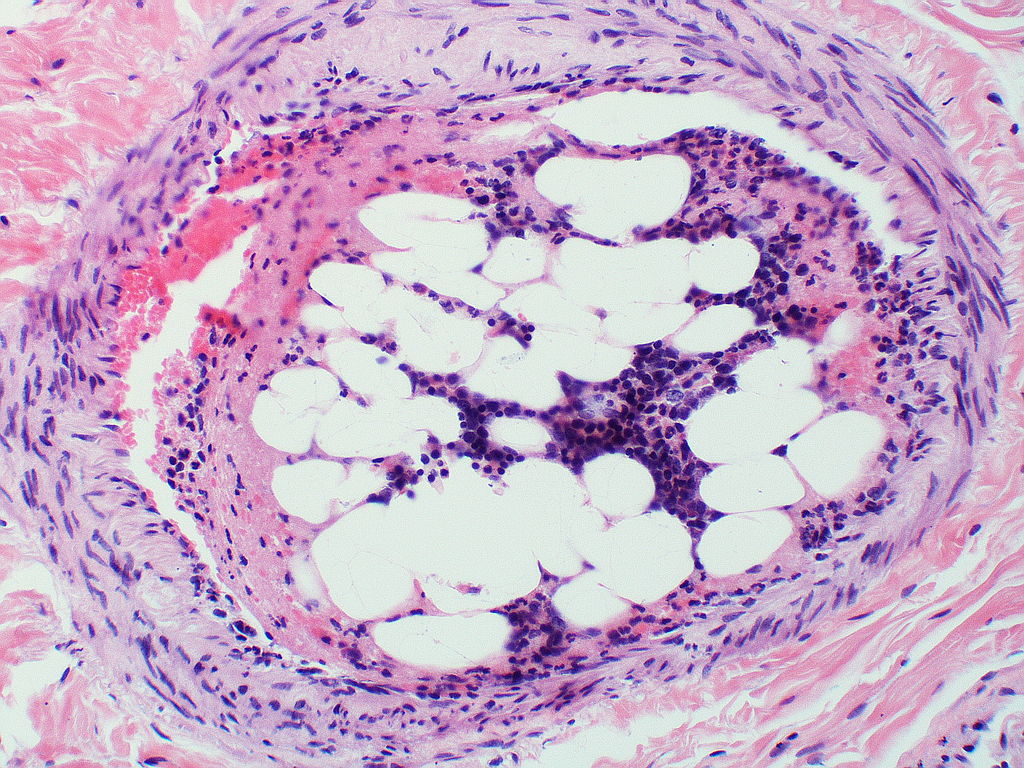
Yale Rosen from USA, CC BY-SA 2.0, via Wikimedia Commons
One challenge which is often overlooked in biomedical research, is the choice of model to use in studies. Trying to investigate the behaviour of cells is like tasting soup: you cannot capture the behaviour of a cell in situ with its neighbours by studying that cell in isolation, in the same way you cannot capture the full breadth of a soup’s flavour by simply eating potatoes on their own.
Cells are surrounded by a network of different cell types around them—those that support it and ensure its normal functioning (called ‘the stroma’), as well as a source of stem cells which replenish those cells that have been lost to wear and tear.
Blood vessels infiltrate most tissues to deliver vital components such as amino acids and glucose, and during any kind of tissue damage or dysfunction, immune cells will invade to patch up (or sometimes, amplify) the damage.
So, the models we use in biomedical research are never ideal, but broadly speaking we can divide them into in vivo, which are living models such as animals, and in vitro, which consist of cells in Petri dishes and test tubes.
Both of these have their disadvantages. Cell organisation in mice will undoubtedly be different from humans, and in vitro models fail to capture those complex cell-cell interactions which occur in tissues.
One new class of model being investigated is the ‘organoid’.
These are tiny, 3D tissues that scientists grow from stem cells, and which can grow to maintain a variety of cell-types, whilst maintaining a particular structure. Although not perfect by any standard, these organoids can be developed to mimic aspects of tissues that researchers want to investigate.
Recently, a team of researchers from the University of Birmingham and the University of Oxford developed a technique to produce organoids that mimic bone marrow.
They used induced pluripotency stem cells (these are stem cells which are made by artificially switching on certain genes in fully differentiated adult cells) and cultured these so they would eventually develop into endothelial and haematopoietic cells. Endothelial cells line blood vessels, and haematopoietic stem cells give rise to all the normal cellular components of blood, such as leukocytes, red blood cells and platelets.
These endothelial and haematopoietic precursors were then grown in hydrogels which contained collagen and Matrigel matrix, two important components for the development of linkages between cells in a tissue. They also added VEGF-C, a signalling protein that promotes the development of blood vessels in bone marrow, which are important in allowing communication with the blood.
As well as understanding the methodology, it’s also important to consider how the scientists knew that their model was effective.
The scientists looked at the RNA of single cells in the organoid and found that it was similar to that of human bone marrow cells, which suggests their behaviour is also similar.
Importantly, the bone marrow organoids were found to be effective models for conditions such as bone marrow fibrosis, which causes extensive scarring during haematological cancers, such as acute leukaemia.
It was found that addition of a protein which increases fibrosis, known as TGF-beta, led to a dose-dependent increase in fibrosis in the organoids, and that inhibitors of TGF-beta could block this fibrosis. Even more crucially, they found that when tumour cells from patients were grown in these organoids, the researchers could test different inhibitors of fibrosis and assess their efficacy. T
his personalised treatment could ensure that patients receive the most effective drug from the start, and this may improve patient outcomes due to earlier optimisation of drug choices.
This new research has direct implications for the treatment of haematological cancers, but perhaps the method they use for developing bone marrow organoids could be extrapolated to develop organoids for other cancers as well.
Organoids are especially important in cancer research because of the extent to which cancerous cells alter their local microenvironment to further their own growth. As the role of the cancer microenvironment becomes even clearer, the need for humanised, 3D models such as organoids will only become greater.