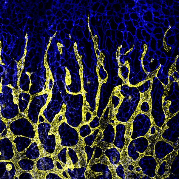
Developing lymphatic vessels in dermal tissue from a mouse. Maria Rondon, CC BY 4.0, via Wikimedia Commons
The lymphatic system is an intricate and often under-appreciated network of vessels responsible for maintaining fluid levels within the body and for transporting cells of the immune system around the body.
Until now, it was believed that human bone and bone marrow was devoid of any lymphatic vasculature. New research conducted by the MRC Human Immunology unit at Oxford has not only revealed the presence of lymphatic vessels within human bone but has demonstrated its crucial role in bone cell regeneration after injury.
Lymph vessels circulate a clear fluid known as lymph around the body, transporting cellular debris, bacteria and proteins as well as any extra fluid from our tissues or blood stream. As lymph passes through lymph nodes, anything harmful is filtered out and removed from the body as urine. As such, swollen lymph nodes are a common symptom of infection and can indicate the body is fighting a foreign pathogen.
Whilst lymphatic tissues are found in almost every organ of the body, a small number of sites are thought to lack any lymphatics. This includes the brain, eye, and until now, bone. Studying the intricacies of blood vessel organisation in bone has been met with difficulty in the past, with the calcified hard tissue of bone being difficult to access and image.
The research, published in Cell, devised a method using light-sheet imaging to visualise intact bones at high resolution to show that lymphatic vessels do in fact exist within the bone. This 3D imaging technique works by illuminating a very thin sample of bone and observing its fluorescent emission from the perpendicular plane. This is then combined with an immunostaining technique, which uses fluorescent antibodies specific for several different lymphatic vessel markers. The results revealed a positive result for antibody binding to these markers within the bone samples, hence confirming the presence of intact lymph vessels.
The study then went on to identify the exact role of lymphatics within bone. They showed that upon injury to bone, the signalling molecule IL6 triggers the expansion of these lymphatic vessels and that this expansion is accompanied by an increase in the number of bone marrow stem cells. This population of stem cells can then reconstitute any injured bone marrow cells.
This finding is of particular therapeutic importance as it offers the potential to target lymphatics in bone and blood regeneration, or to even promote healing in fracture repair. As Dr Anjali Kusumbe, Group Leader of the Tissue and Tumor Microenvironments Group at the MRC Human Immunology Unit, who led the research said: ‘these findings are very fundamental, opening doors for understanding the impact of bone lymphatics on the immune system and their role in bone and blood diseases.’
Moreover, the study suggested that impaired lymphatic signalling may account for the reduced capacity for repair and regeneration in aged bones. Dr Junyu Chen, a co-first author of the study said: ‘Aging is associated with diminished capacity for bone repair, and our findings show that lymphatic signalling is impaired in aged bones. Remarkably, the administration of young lymphatic endothelial cells restores healing of aged bones, thus providing a future direction to promote bone healing in elderly.’
Whilst this discovery represents a large step forward in our understanding of the lymphatic system, it also presents many questions, paving the way for future research endeavours. Elucidating the exact role of these lymph vessels in disease states such as rheumatoid arthritis, and not just in the context of physiological wound healing, will be required if these findings are to be implemented therapeutically.