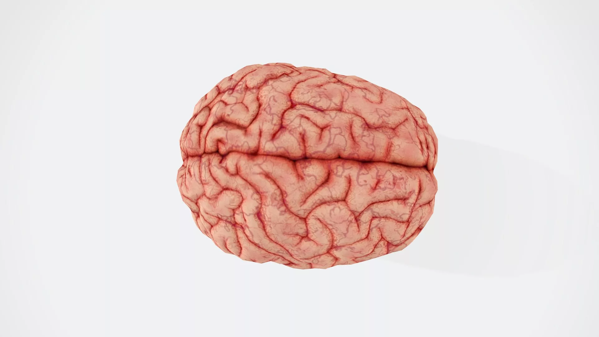
The human brain is the most complex organ in nature. This complexity has historically been hard to organise however, recent work is aiming to create an atlas of the human brain. Photo credit: BUDDHI Kumar SHRESTHA via Unsplash
Contemporary neuroscience as we know it today owes much of its origins to the drawings of Spanish histologist Santiago Ramón y Cajal. The father of modern neuroscience, Cajal produced countless intricate sketches depicting the individual cells that make up the brain. At a time when scientists believed the brain to be a continuous tangle of fibres, akin to a serpent or a labyrinth, the discovery of these discrete units or neurons won Cajal the Nobel prize in 1906.
Over a century later, neuroscience is again rediscovering its artistic streak. Indeed, a recent special edition of Science presents a collection of twelve papers attempting to synthesise individual profiles of neurons into an “atlas” of the human brain. A further nine papers are published in its spin-off journals Science Advances and Science Translational Medicine. The work emerges from the Brain Initiative Cell Census Network (BICC)–a consortium of research centres across the US and Europe organised by the National Institute of Health (NIH). Ambitiously, the network aims to characterise and map every class of neuron in the human brain.
A detailed “atlas” of the brain’s cellular composition, therefore, could prove invaluable in demystifying its complexity.
This atlas could offer a cellular insight into the marvellous intricacy of our brains—the most complex objects in the known universe. The brain is composed of billions of neurons and supporting (non-neuronal) glial cells organised into overlapping regions, each responsible for remarkably diverse cognitive processes. For example, the occipital lobe at the back of the brain is broadly responsible for vision whilst the frontal lobes—aptly named due to their location at the front of the brain—govern higher functions like decision making. This anatomical division of labour, which neuroscientists refer to as functional specialisation, is central to the complexity of the brain. A precise understanding of the cells that constitute the human brain will help illuminate how this functional specialisation is implemented. After all, regional differences in how information is processed are likely to be reflected in the processing units that make up each structure. A detailed “atlas” of the brain’s cellular composition, therefore, could prove invaluable in demystifying its complexity.
A transcriptomic revolution
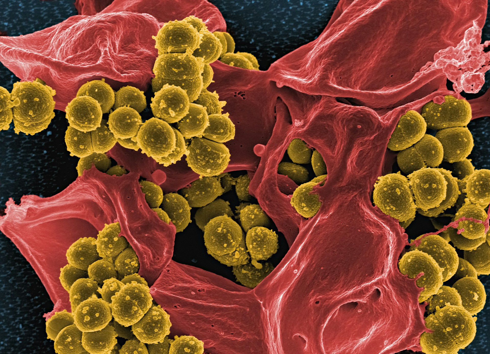
The development of technology for genetically profiling individual cells—previously only achieved with efficiency in animal models—now makes this a feasible goal. Indeed, many of the papers in this special edition use single-nucleus RNA sequencing to systematically survey cells across the human brain. Strands of RNA act as chemical messengers within cells. They carry instructions on how to make proteins from genes held in the nucleus to the ribosomes where the proteins are made. Analysing the RNA transcripts the cell holds, therefore, reveals the proteins that are being made within it. Given that proteins do much of the chemical work inside a cell, this largely determines the kind of cell that it is. Researchers are thus using RNA sequencing to generate a genetic “profile” of each cell in the brain.
One study in the edition emerging from the Karolinska Institutet, Stockholm, and the Allen Institute for Brain Sciences, Seattle, attempted to synthesise these profiles into the cell “types” that populate the brain. Using a statistical technique called cluster analysis, they grouped individual neurons together based on similarities in their RNA transcripts. This yielded over 3,000 distinct types of cells: a quite staggering level of microscopic diversity. For context, not long ago, the entire human body—brains included—was thought to consist of just 300 types of cells. These 3,000-something types could then be grouped into 461 clusters and 31 “superclusters”, based on differing statistical levels of similarity in their RNA transcripts. The researchers present these clusters as a genetic “key” for the types of cells that populate the human brain.
Other groups joined in the project of atlas building, mapping where these neuronal types exist in the brain. This produced an intriguing set of findings. Most notably, different brain areas contained the same major classes of cell types despite carrying out drastically different cognitive functions. The brain lacks cells that exist uniquely in one region or another. The Stockholm-Seattle group identified a supercluster termed “splatter neurons”, containing neurons that featured prominently in nearly every area of the brain. Variations in the cognitive processing carried out by each of these regions, therefore, were not underpinned by discrete differences in the unique cell types they possess. Instead, functional specialisation was better explained by the relative proportions of each cell type found in different brain regions.
The ratio of inhibitory to excitatory neurons in the primary visual cortex, another research team found, was more than double that of other cortical areas. Excitatory neurons promote activation in downstream cells when they fire, whereas inhibitory neurons decrease the likelihood of such activation. The primary visual cortex carries out the first cortical processing of visual information and is specialised for pattern recognition. It receives input directly from the retina via the lateral geniculate nucleus. Hence, it makes sense that inhibitory neurons are so prominent in this area. Their role is critical in down-regulating neural activity in the region, which is constantly stimulated by sensory input. Overall, regional differences in cognitive processing seem to be reflected in the relative proportion of each cell type each brain region possesses.
This data suggests that the diversification of regional function evolved not through the generation of new cell types but via small genetic variations within them. In altering the relative propensity of existing cell types to pop up in different regions of the brain, these minor genetic adjustments can create the circuitry distinctive of human cognition.
Another study from the edition concurs. They compared the RNA transcripts of cells from a region critical for language comprehension—the medial temporal gyrus (MTG)—across humans and four non-human primate species. Transcriptomic profiles in the MTG appeared to be largely conserved across 40 million years of primate evolution despite language only surfacing in the human brain. However, the relatively sparse areas of the genome that do show signs of evolutionary selection in humans had an outsized impact: the information contained in these genetic regions is expressed in ways unique to humans, to a much greater extent than other areas of DNA. Clues to the human specialisation of the MTG for language, therefore, are likely to reside in these relatively minor genetic adjustments. Intriguingly, the brain cells that seem to be particularly affected by the expression of genetic information near these regions are glia: the non-neuronal bodies that perform a range of functions in the brain, from regulating the flow of information between neurons to pruning links between them. The cognitive specialisation of the human brain, therefore, may derive from relatively few genetic changes to the scaffolding that holds up neuronal circuitry.
These are remarkable findings: what gives human brains their humanness may lie in the microscopic details of their architecture. But they also illustrate the danger of relying solely on the RNA transcripts of a cell in determining its identity. Sometimes, interesting data can get lost in the inherent noise of transcriptomic profiles. Although the above study comparing primate species found signs of evolutionary selection in the human transcriptome, these differences were relatively minute. Language is a unique feature of the human brain, yet transcriptomic profiles in the human MTG showed remarkable similarity to non-human primates. Rather, the crucial point of interest lay in how the genes near these regions were differentially expressed between species and what this meant for cells in that area of the brain. Transcriptomic profiles mark a great starting point, but they need to be updated as data from new modalities is obtained. An atlas based on transcriptomes alone may overlook important details.
Towards a more holistic understanding
The utility of a multimodal approach is demonstrated by another paper in the edition from researchers at the University of California, San Diego (UCSD). They attempted to characterise cell types, not directly by their messenger RNA, but by differences in the way the DNA in their chromosomes was packaged in chromatin formations. These structures serve to bundle long DNA molecules into more compact structures in ways that allow or forbid RNA messengers access to certain regions of the genome. In this sense, chromatin formations influence which genes are transcribed and, hence, expressed in the chemical functions of the cell.
Analysing these formations yielded 107 recognisable patterns. Intriguingly, these chromatin types could shed light on the cellular basis of neuropsychiatric disorders such as depression and schizophrenia. Previous studies have identified risk variants of genes, the possession of which increases the likelihood that the holder suffers from a given neuropsychiatric illness. The researchers at UCSD compared the locations of pieces of DNA that control gene transcription, and are known to have these disease-associated variants, with the different classes of chromatin formations they uncovered. This revealed the formation types that allowed “risky” transcriptions to occur and, hence, the cells that are implicated in different neuropsychiatric disorders. The researchers were able to correlate these cell types with nineteen different conditions, including depression, schizophrenia, and various forms of addiction. These correlations would not have emerged by analysing RNA transcriptomes alone.
Where to go from here?
So herein lies the challenge of “atlasing” the brain: neuroscientists must continually refine and annotate their cellular taxonomies, such that mapping where these types of neurons exist in the brain will tell us all we want to know about cognitive functioning. This is an immense task that is by no means complete. Scientists need ever new ways to query how “real” their classifications are, says Nathan Gouwens—a computational neuroscientist at the Allen Institute for Brain Sciences.
And exciting new methodologies are continuing to emerge. In a special edition packed with cutting-edge science, some of the most impressive findings came from researchers utilising Patch-sequencing—a technique facilitating simultaneous recordings of the transcriptome, electrical activity, and shape of human brain cells surveyed in post-mortem or after neurosurgery. A researcher navigates their pipette to a cell and adds suction to gain electrical access. Measuring the amount of voltage that passes through the cell at a fixed amount of current, and vice-versa, gives the researcher insight into its electrical behaviour. Throughout these electrophysiological recordings, dye is infused into the cell to yield data about its shape. As a result, researchers are beginning to annotate their taxonomies with stunning colour images that Cajal himself would admire. The pipette is then used to extract the cell’s nucleus for transcriptomic analysis. With Patch-seq, a lab can assess how similar or dissimilar individual cells are to neighbours in terms of their transcriptome, electrical activity, and shape. This could uncover new realms of cellular diversity in the brain.
Indeed, researchers are finding that cells classically lumped together actually belong to different cell types when the brain is studied in greater multimodal detail. This can help validate and refine the exciting results emerging from large-scale transcriptomic assays of the brain. Even within transcriptomic cell types, one paper in the edition found the shape of neurons can differ drastically–and these morphological differences showed strong coherence with the electrophysiological features of the neurons. Appreciating these features will provide much-needed “biological context” to genetic taxonomies of the cells that populate the brain, says Tibor Harknay, who has a double appointment at the Medical University of Vienna and the Karolinska Institutet.
If a cellular atlas is to help explain the remarkable feats of the brain, therefore, its “key” must be annotated with more than just the genetic properties of cells.
After all, we want to know the role that cellular diversity plays in the functioning of the brain: the ways that different cells represent information in their electrical activity and transmit it to their neighbours by virtue of their unique shapes. This will help explain “how” different transcriptomic cell types underpin distinct cognitive functions; or “why” they are implicated in certain neuropsychiatric disorders. A key project for neuroscience is investigating the rules governing variations in these functional properties. If a cellular atlas is to help explain the remarkable feats of the brain, therefore, its “key” must be annotated with more than just the genetic properties of cells.
Nevertheless, something exciting is beginning to emerge. Since Cajal’s remarkable sketches first began to unveil the immense diversity of cells found in the brain, neuroscience has been challenged to understand how this diversity underpins the functional complexity of the brain. Today, exciting new technologies in single-cell analysis make meeting this immense challenge a reality. Yet we should be conservative about the conclusions we draw at this stage; even a brief look at history reveals how inaccurate the early attempts of cartographers can be. Science has published a good start. But the journey of neuroscience into the cellular complexity of the brain is a fledgling one.